
Viruses are microscopic infectious agents that comprises of genetic material (DNA or RNA) encapsulated inside a protein coat. It infects the living cells of an organism in order to replicate itself which usually results in various diseases. This whole replication process involves the attachment and penetration of the virus to the host cell, replication of viral genetic material, the maturation of new viral particles and finally, their release from the host cell.
The most common example of diseases caused by viruses are COVID-19, AIDS, measles and chickenpox. However, the presence of antiviral agents can inhibit the infection or replication of viruses in living cells. What if we say you can have a visual image of virus replication occurring in cells?
We are delighted to announce that TECOLAB is now offering a creatively developed technology that provides real time visualization of virus infection and replication in living cells with the help of our collaborative partner.
How does this technology work?
This technology allows the binding of fluorescent proteins to short sequences of viral DNA, resulting in the exhibition of fluorescent spots that correspond to the position of virus genetic material. These spots can be visualized and quantified under a microscope. The tagged sequence can be easily inserted into the DNA of interest (plasmid, gene, virus, viral vector) without disrupting the metabolism of DNA and virus production. This allows for precise tracking of specific cells or DNA sequences in living organisms and can provide valuable insights into various biological process. In order to ensure the highest quality of visualization, we offer high content imaging, high resolution imaging, and light sheet microscopy. For instance, this technology can be used to rapidly assess various customer parameters for disinfection efficiency including on highly pathogenic viruses like SARS-CoV-2.
Check out some images below on how the visualization of viruses will look!
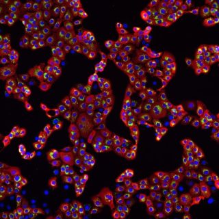
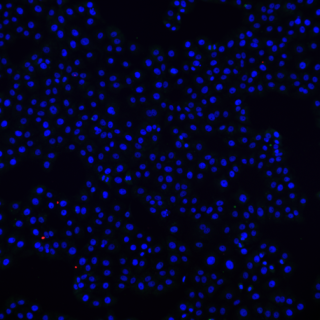
Visuals of transition of UV-C light effectively killing human infectious adenoviruses in the cell after 10 minutes as there is no presence of nucleocapsid protein (red) or SARS RNA (green) after UV-C exposure.
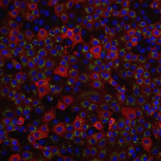
Blue: Nuclei of cell , Green: SARS-CoV-2 RNA , Red: SARS -CoV-2 nucleocapsid protein


Before UV-C exposure, 0 min
After UV-C exposure, 10 mins
Magnified view of SARS-CoV-2 infected cells
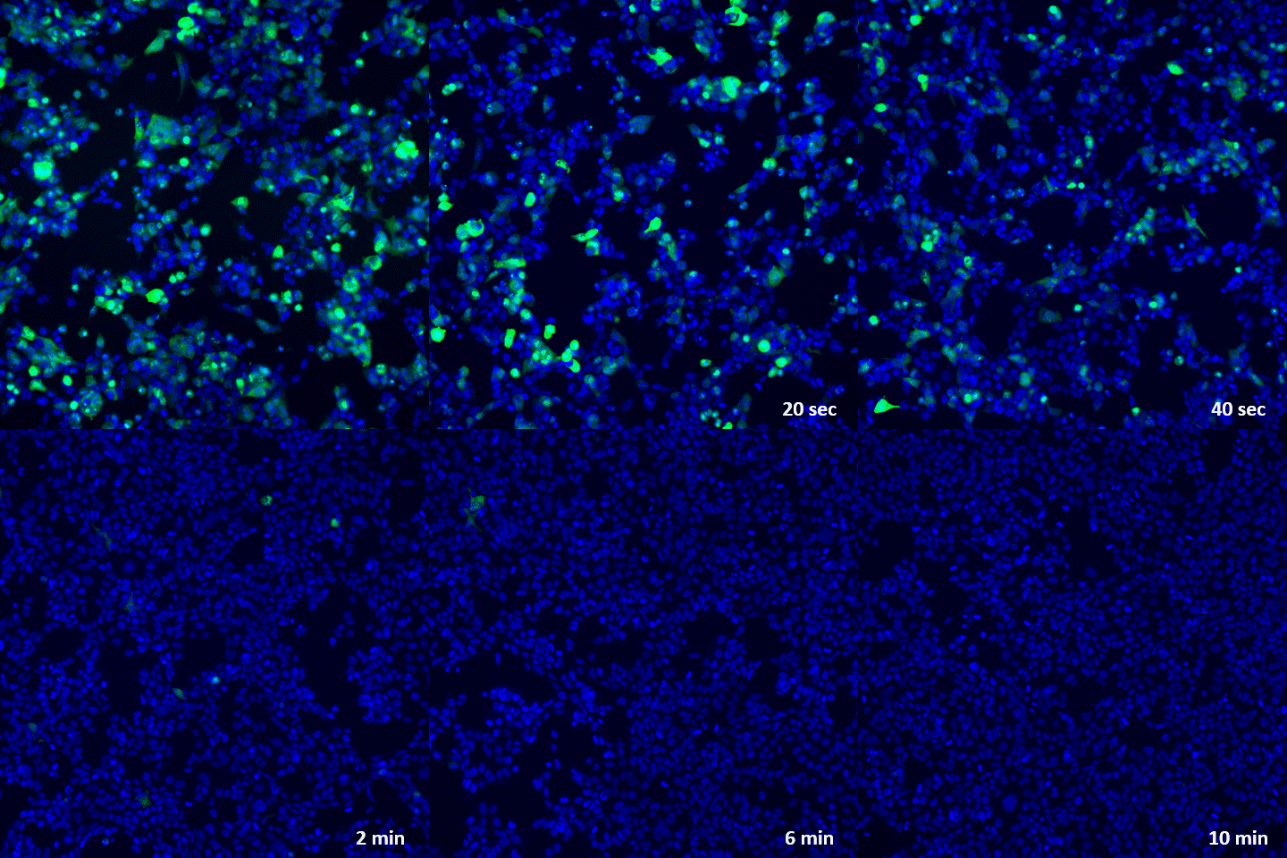
The animated GIF below demonstrates the progressive killing of the virus from 0 minutes to 6 minutes, with only the nucleus of the cell remaining at the end. The green fluorescence observed in the animation indicates the virus DNA (hAdv5)

What viruses can be used with this technology?
Technically, this technology can be used in any viruses with double-stranded DNA but we have a list of readily available viruses for you to choose from below. If you are interested in any viruses that are not listed, let us know and we will work something out for you.
| Type of Viruses | |
| Herpes virus | Varicella-zoster virus (VZV) |
| Vaccinia virus | Feline calicivirus |
| Retrovirus (eg. HIV) | Parvovirus |
| Adenovirus | African swine fever virus (ASFV) |
| Epstein-Barr virus (EBV) | Respiratory syncytial virus (RSV) |
| SARS-CoV-2 | Baculovirus |
| Influenza virus | Rhinovirus |
What are the services this technology can offer?
Revolutionize the marketing of your disinfection devices by providing photos and videos of the effectiveness of your products using this advanced fluorescent virus technology!
Up to now, log reduction or killing rate is a common tool in displaying the effectiveness of disinfectants. While this information is vital and is widely recognized, it would look even better and compelling when accompanied with a clear before-and-after-picture or a video to demonstrate inactivation of viruses during the disinfection process. This can be effective in industries such as healthcare, where preventing the spread of infections is critical.
Drug discovery is the identification of chemical compounds that may have the ability to become therapeutic agents for various diseases. These fluorescent viruses become an important resource for antiviral research and development as they can be utilized in high-throughput imaging and screening. In addition, the integration of HCS microscopy and detection of algorithms enables the rapid and reliable discovery of new antiviral candidates. With this technology, we will be able to quickly establish the complete pre-clinical antiviral development process, for example, by identifying their mechanism of action.
By incorporating fluorescent proteins into oncolytic viruses or other cancer therapeutics, this technology enables real-time tracking of viral replication and distribution within cells. This allows researchers to monitor the efficacy of potential cancer therapeutics and gain insights into their mechanisms of action.
One advantage of this technology is its ability to provide high-resolution imaging of oncolytic viruses within cells. By tracking viral replication and distribution in real time, researchers can gain insights into how the viruses are interacting with their target cells and how they may be affecting tumor growth and progression. This information can be used to identify promising candidates for further development and optimize treatment regimens.
Various types of research can benefit through this technology such as the development of inactivation solutions testing for antivirals, and inventing decontamination tools (eg. UV or ozone) on different types of mediums such as cell lines, live human skin explants, surfaces (eg. stainless steel, tissues, masks, etc.).
Contact us here to start your journey with us using this all-new fluorescent virus technology.